Page 80 - Kutnar, Andreja, et al., eds., 2015. Proceedings of the 1st COST Action FP1307 International Conference - Life Cycle Assessment, EPDs, and modified wood. University of Primorska Press, Koper.
P. 80
swax
impregnation
increased
the
MOE
of
beech
and
poplar
wood.
Unimpregnated
beech
and
poplar
samples
decomposed
completely
during
the
18
months
of
soil
contact.
The
damage
of
the
impregnated
samples
was
markedly
lower.
This
was
confirmed
by
the
MOE
measurements,
which
showed
remarkable
remaining
MOE
of
the
impregnated
samples
after
soil
exposure
(Fig.
1).
The
impregnation
improved
the
wood’s
resistance
against
wood
decaying
organisms,
and
higher
DPS
resulted
in
less
of
a
decrease
in
MOE
than
in
samples
with
lower
DPS.
Figure
1:
MOE
of
poplar
and
beech
samples
in
the
investigation
periods.
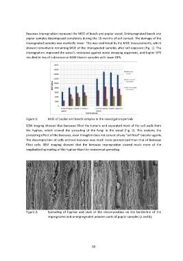
SEM
imaging
showed
that
beeswax
filled
the
lumens
and
separated
most
of
the
cell
walls
from
the
hyphae,
which
slowed
the
spreading
of
the
fungi
in
the
wood
(Fig.
2).
This
explains
the
protecting
effect
of
the
beeswax,
even
though
it
does
not
consist
of
any
“artificial”
biocidal
agents.
The
decomposition
of
cells
without
beeswax
was
much
more
pronounced
than
that
of
beeswax
filled
cells.
SEM
imaging
showed
that
the
beeswax
impregnation
slowed
much
more
of
the
longitudinal
spreading
of
the
hyphae
than
the
transversal
spreading.
Figure
2:
Spreading
of
hyphae
and
start
of
the
decomposition
on
the
borderline
of
the
impregnated
and
unimpregnated
wooden
parts
of
poplar
samples
(a
and
b).
68
impregnation
increased
the
MOE
of
beech
and
poplar
wood.
Unimpregnated
beech
and
poplar
samples
decomposed
completely
during
the
18
months
of
soil
contact.
The
damage
of
the
impregnated
samples
was
markedly
lower.
This
was
confirmed
by
the
MOE
measurements,
which
showed
remarkable
remaining
MOE
of
the
impregnated
samples
after
soil
exposure
(Fig.
1).
The
impregnation
improved
the
wood’s
resistance
against
wood
decaying
organisms,
and
higher
DPS
resulted
in
less
of
a
decrease
in
MOE
than
in
samples
with
lower
DPS.
Figure
1:
MOE
of
poplar
and
beech
samples
in
the
investigation
periods.
SEM
imaging
showed
that
beeswax
filled
the
lumens
and
separated
most
of
the
cell
walls
from
the
hyphae,
which
slowed
the
spreading
of
the
fungi
in
the
wood
(Fig.
2).
This
explains
the
protecting
effect
of
the
beeswax,
even
though
it
does
not
consist
of
any
“artificial”
biocidal
agents.
The
decomposition
of
cells
without
beeswax
was
much
more
pronounced
than
that
of
beeswax
filled
cells.
SEM
imaging
showed
that
the
beeswax
impregnation
slowed
much
more
of
the
longitudinal
spreading
of
the
hyphae
than
the
transversal
spreading.
Figure
2:
Spreading
of
hyphae
and
start
of
the
decomposition
on
the
borderline
of
the
impregnated
and
unimpregnated
wooden
parts
of
poplar
samples
(a
and
b).
68